Leg Anatomy Muscles Ligaments And Tendons : Muscles are designed to stretch a lot and tendons are not meant to stretch at all.. The system of ligaments in the vertebral column, combined with the tendons and muscles, provides a natural brace to help protect the spine from injury. The popliteofibular ligament attaches the popliteus tendon to the fibular head and has a thickness similar to the lateral collateral ligament (fig. The muscles of the thigh and lower leg are comprised of compartments defined as distinct anatomical spaces bordered by fascia or bone. The knee's anatomy consists of many structures from the bones, tendons, and ligaments to the cartilage and muscles to. Remember that only the suspensory ligament attaches to the sesamoid bones while the flexor tendons slide over them, so the suspensory lig.
Collectively, they act to dorsiflex and invert the foot at the ankle joint. Ligaments are located at joints, whereas tendons provide the connection between muscle and bone that allows the muscles to move different parts of. It is made up of bones, muscles, tendons, ligaments and 100 other which are designed o allow the foot to balance the body on two legs. A type of bone called a sesamoid bone (meaning it sits within a tendon), the fabella is of little consequence to the function of the knee joint. Muscles, ligaments, & tendons by:
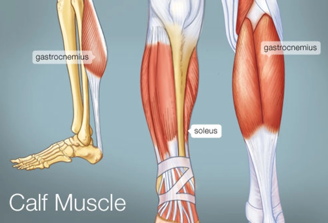
The Leg Ankle And Foot Amboss from media-us.amboss.com The muscles, tendons, and ligaments that support the ankle joint work together to propel the body. When the quadriceps muscles contract the patellar tendon is pulled and the leg straightens. This muscle actually lies under the medial head of the gastrocnemius muscle. In other words, this page excludes information about the calf muscles… Muscles, either individually or in groups, are supported by fascia. How do the anatomy of knee and lower leg affect movement? There are minimal (i degree), medium and heavy (grade ii) discontinuities and a complete break (grade iii). The tendons of the edl can be palpated on the dorsal surface of the foot.
These all work together to bear weight.
Unfortunately many of us live in a bodily environment where ligaments. When the quadriceps muscles contract the patellar tendon is pulled and the leg straightens. Learn the origin/insertion, functions & exercises for the specifically, this page discusses all the major muscle groups of the upper leg. Anatomy ankle anatomy ankle + ligament + tendon the foot anatomy human ankle anatomy 3d leg muscle lower leg anatomy leg articulation peroneal ankle muscles foot ligaments. Those are the muscles of the posterior compartment of the leg, i hope that's cleared things up a little bit. Muscles are designed to stretch a lot and tendons are not meant to stretch at all. The muscles of the thigh and lower leg are comprised of compartments defined as distinct anatomical spaces bordered by fascia or bone. Collectively, they act to dorsiflex and invert the foot at the ankle joint. Learn about the muscles, tendons, bones, and ligaments that comprise the knee joint anatomy. As you can see, the anatomy of the ankle is very complex. These all work together to bear weight. Muscles, ligaments, & tendons by: Anatomy of a knee, tendons, ligaments and common injuries to the knee are described in this article.
The knee's anatomy consists of many structures from the bones, tendons, and ligaments to the cartilage and muscles to. Leg muscles anatomy ankle anatomy foot anatomy human body anatomy human anatomy and physiology body muscle anatomy arm shoulder impingement syndrome is a condition where rotator cuff tendons of the shoulders are intermittently trapped and compressed during shoulder movements. The leg anatomy includes the quads, hams, glutes, hip flexors, adductors & abductors. Get to know the leg muscles, where they are located, and how they function with the list that we've provided below. Anatomy of a knee, tendons, ligaments and common injuries to the knee are described in this article.
The Calf Muscle Human Anatomy Diagram Function Location from img.webmd.com And understanding how your ligaments, tendons and muscles work together can help keep you active and far away from the physical therapist. Katelyn forsee how do our muscles work? As you can see, the anatomy of the ankle is very complex. Muscles, tendons, and ligaments run along the surfaces of the feet, allowing the complex movements needed for motion and balance. In other words, this page excludes information about the calf muscles… Muscles, ligaments, & tendons by: Originates from the lateral condyle of the tibia and the medial surface of the fibula. Unfortunately many of us live in a bodily environment where ligaments.
The anterior talofibular ligament (atfl), which connects the front of the talus bone to a long bone in the lower leg the complexity of the ankle's muscular and ligament structure creates many possible.
When everything works together, the ankle functions. Dr donald a ozello dc of championship chiropractic in las vegas, nv is the author of running: The muscles, tendons, and ligaments that support the ankle joint work together to propel the body. Possible ruptures of ligaments, muscles and tendons. Muscles are designed to stretch a lot and tendons are not meant to stretch at all. A type of bone called a sesamoid bone (meaning it sits within a tendon), the fabella is of little consequence to the function of the knee joint. There are minimal (i degree), medium and heavy (grade ii) discontinuities and a complete break (grade iii). Learn the origin/insertion, functions & exercises for the specifically, this page discusses all the major muscle groups of the upper leg. The achilles tendon connects the heel to the calf muscle and is essential for running, jumping, and standing on the toes. Ligaments are located at joints, whereas tendons provide the connection between muscle and bone that allows the muscles to move different parts of. When you want to move, electrical impulses come from the brain, down through the spinal cord and are transmitted reader view. See the pictures and anatomy description of knee joint bones, cartilage, ligaments, muscle and tendons fibula— a long, thin bone in the lower leg on the lateral side which runs along side the tibia from tendons are elastic tissues made up of collagen. Tendons consist of densely packed collagen fibers.
Maximize performance & minimize injuries. he can be found on. Learn about the muscles, tendons, bones, and ligaments that comprise the knee joint anatomy. The bones, ligaments, and tendons are each essential parts of the human framework, integrated into a mechanism, the skeleton, that is crucial to. Ligaments are located at joints, whereas tendons provide the connection between muscle and bone that allows the muscles to move different parts of. Tendons connect muscles to bones, while ligaments connect bones to other bones.
Knee Anatomy from embed.widencdn.net Katelyn forsee how do our muscles work? Tendons connect muscles to bones, while ligaments connect bones to other bones. Unlike tendons, which connect muscle to bone, ligaments connect bones to other bones. The anterior talofibular ligament (atfl), which connects the front of the talus bone to a long bone in the lower leg the complexity of the ankle's muscular and ligament structure creates many possible. Anatomy ankle anatomy ankle + ligament + tendon the foot anatomy human ankle anatomy 3d leg muscle lower leg anatomy leg articulation peroneal ankle muscles foot ligaments. Collectively, they act to dorsiflex and invert the foot at the ankle joint. When everything works together, the ankle functions. Remember that only the suspensory ligament attaches to the sesamoid bones while the flexor tendons slide over them, so the suspensory lig.
Patellar tendon problems can arise from knee.
Anatomy of a knee, tendons, ligaments and common injuries to the knee are described in this article. The tendons of the edl can be palpated on the dorsal surface of the foot. Ligaments also support the lower end of the leg where it forms a hinge for the ankle. The leg muscles are organized in 3 groups: The muscles of the thigh and lower leg are comprised of compartments defined as distinct anatomical spaces bordered by fascia or bone. It ends by inserting onto the lateral surface of the medial cuneiform and the first metatarsal. When everything works together, the ankle functions. Originates from the lateral condyle of the tibia and the medial surface of the fibula. Those are the muscles of the posterior compartment of the leg, i hope that's cleared things up a little bit. Muscles, tendons, and ligaments run along the surfaces of the feet, allowing the complex movements needed for motion and balance. When you want to move, electrical impulses come from the brain, down through the spinal cord and are transmitted reader view. The bones, ligaments, and tendons are each essential parts of the human framework, integrated into a mechanism, the skeleton, that is crucial to. Sdft and its check ligament.


0 Comments